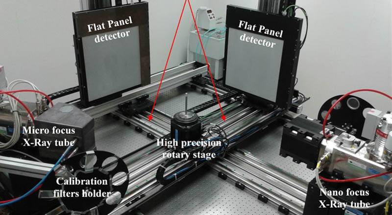
X-ray 3D Computed Tomography (CT) is a nondestructive scanning technology that allows to view and inspect the external and internal structures of an object in 3D space. Computed Tomography works by taking hundreds or thousands of 2D Digital Radiography projections around a 360 degree rotation of an object. Proprietary algorithms are then used to reconstruct the 2D projections into a 3D CT volume, which allows to view, inspect and slice the virtual object, as well as to perform various analyses, such as porosity distribution etc. X-ray 3D Computed Tomography (CT) is a nondestructive scanning technology that allows to view and inspect the external and internal structures of an object in 3D space. Computed Tomography works by taking hundreds or thousands of 2D Digital Radiography projections around a 360 degree rotation of an object. Proprietary algorithms are then used to reconstruct the 2D projections into a 3D CT volume, which allows to view, inspect and slice the virtual object, as well as to perform various analyses, such as porosity distribution etc. The X-ray CT device available at ITAM (Twinned Orthogonal Adjustable Tomograph - TORATOM) comprises of two orthogonal imaging axes and a shared rotational stage. The magnification is adjustable between 1.2x to 150x, allowing thus reaching the effective pixel size from 1 micrometer. The maximum size of the investigated object is limited to 350 mm in diameter (reaching the resolution of approx. 150 micrometers per voxel) and height 800 mm, its maximum weight is 20 kg. The device is equipped with a reflection type 240 kV / 260 W microfocus X-ray tube and with a transmission type 160 kV / 25 W microfocus X-ray tube with the nanofocus mode available. There are scintillation detectors with the matrix of 2048 x 2048 active pixels and 200 micrometers pixel pitch, scintillation detector with the matrix of 1536 x 1944 active pixels and 75 micrometers pixel pitch and photon-counting detector based on TimePix detectors, with 2560 x 2560 active pixels and 55 micrometers pixel pitch. The double-axis design of TORATOM enables comfortable dual-source or dual-energy tomographies in special cases. The double-axis design of TORATOM enables comfortable dual-source or dual-energy tomographies in special cases.
Fields of application
-
Cultural heritage
art, decorative arts, demo anthropologic object, manuscript, mosaics, musical instrument, other, painting, papyrus, sculpture, textile
-
Natural heritage
fossil, mineral, object in formalin, other, shell, skeleton
Materials
-
inorganic
glass, stone, pigment
-
organic
animal parts, wood, paper, textiles
TOOLS
X-ray 3D Computed Tomography (CT) is a nondestructive scanning technology that allows to view and inspect the external and internal structures of an object in 3D space. Computed Tomography works by taking hundreds or thousands of 2D Digital Radiography projections around a 360 degree rotation of an object. Proprietary algorithms are then used to reconstruct...